Understanding the Path to Cancer Development
In this section, you will learn:
- Cancer is a collection of diseases in which some of the body’s cells acquire changes that allow them to grow uncontrollably and spread to other parts of the body.
- Basic research has played a pivotal role in understanding how cancer begins and spreads.
- Changes inside the cell and influences from outside the cell drive cancer initiation and progression.
- Technological advances have accelerated the identification of molecular and cellular mechanisms that drive cancer.
- Integrating the knowledge gained from decades of research into cancer development has fueled the field of precision medicine.
Contents
Precise molecular mechanisms control normal cell growth and division, which are required for development, regeneration, and overall healthy tissue and organ functions. In cancer, these mechanisms go awry, allowing cells to multiply uncontrollably and spread to other parts of the body. Cancer is not one disease, but a collection of related diseases that can affect nearly any part of the body. Throughout the course of cancer development, abnormal or damaged cells acquire distinct traits—known as the “hallmarks of cancer”—that set them apart from normal cells.
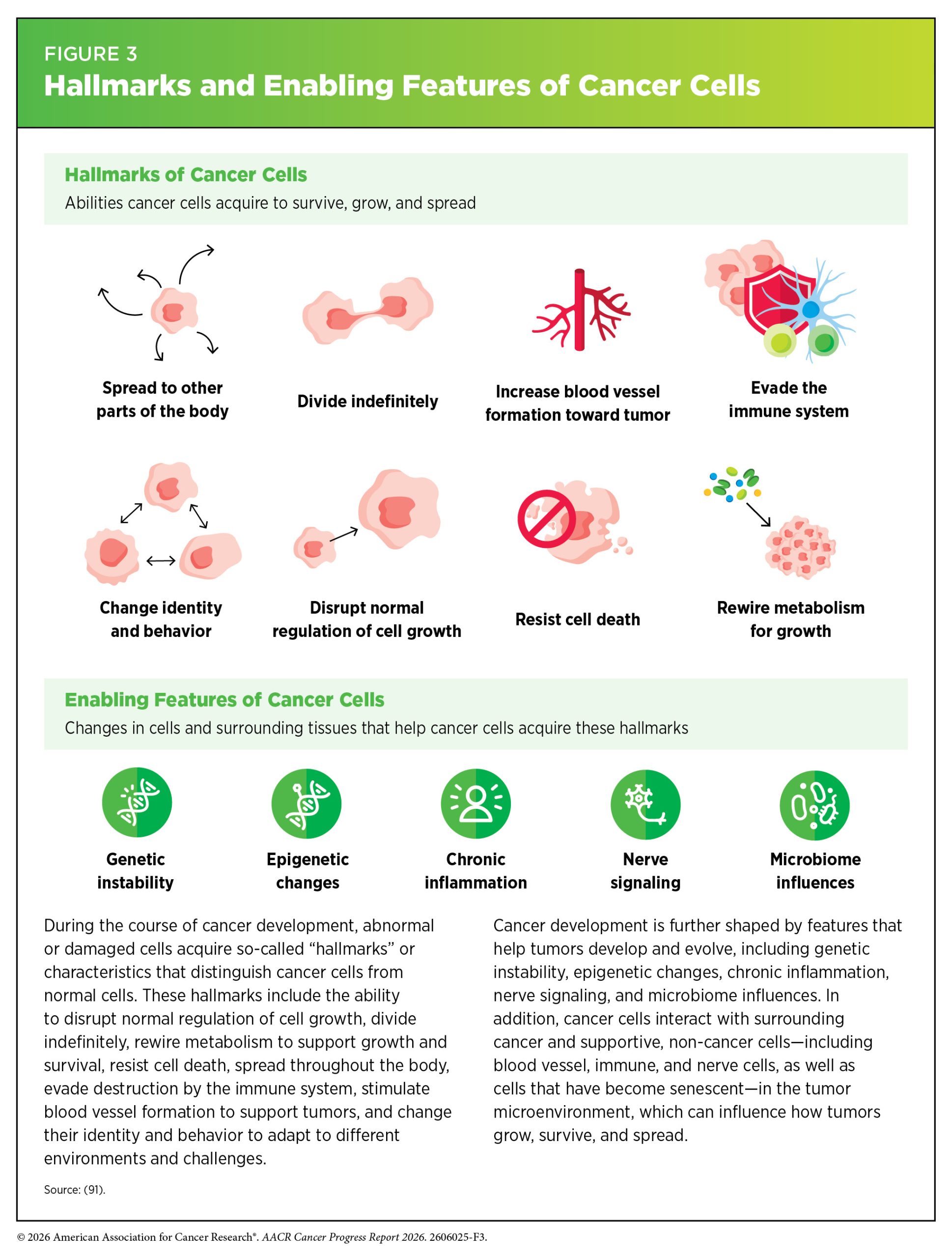
These hallmarks include the ability of cancer cells to divide limitlessly, disrupt normal regulation of cell growth, change identity and behavior, rewire metabolism to support rapid growth, resist cell death, stimulate blood vessel formation, evade the immune system, and leave the tissue of origin and spread to other organs (see Figure 3) (91)Hanahan D (2026) Cell, 189: 2254.. Cancer that has spread to other parts of the body, which is called metastatic disease, is the main cause of most cancer-related deaths.
Generating Knowledge Through Basic Research
Basic research focuses on gaining new knowledge or a deeper understanding of phenomena or observable events. For example, basic research plays an essential role in characterizing normal cell behavior and identifying the changes that drive cancer initiation and progression. The knowledge gained from basic research deepens our understanding of cancer biology and provides a foundation for discovering new ways to target cancer cells, developing more effective treatments, and improving strategies for early detection and prevention. In recognition of the critical role of basic research in improving overall health for all individuals, more than 50 percent of the National Institutes of Health (NIH) budget was allocated to basic research each year from 2003 through 2024, the most recent year for which data are available (92)National Institutes of Health. The Office of Budget. National Institutes of Health FY 2003 – FY 2024 Distribution of Budget Authority Percentages for Basic and Applied Research. Accessed: June 30, 2026..
Discoveries from basic research have transformed cancer care by revealing the fundamental biological mechanisms that drive cancer. For example, studies of cancer-causing viruses beginning in the early 1900s revealed genes that drive cancer development and laid the foundation for many modern targeted therapies (see Basic Research Decoding Cancer’s Complexities). These advances have depended on experimental models that allow researchers to study cancer under controlled conditions.
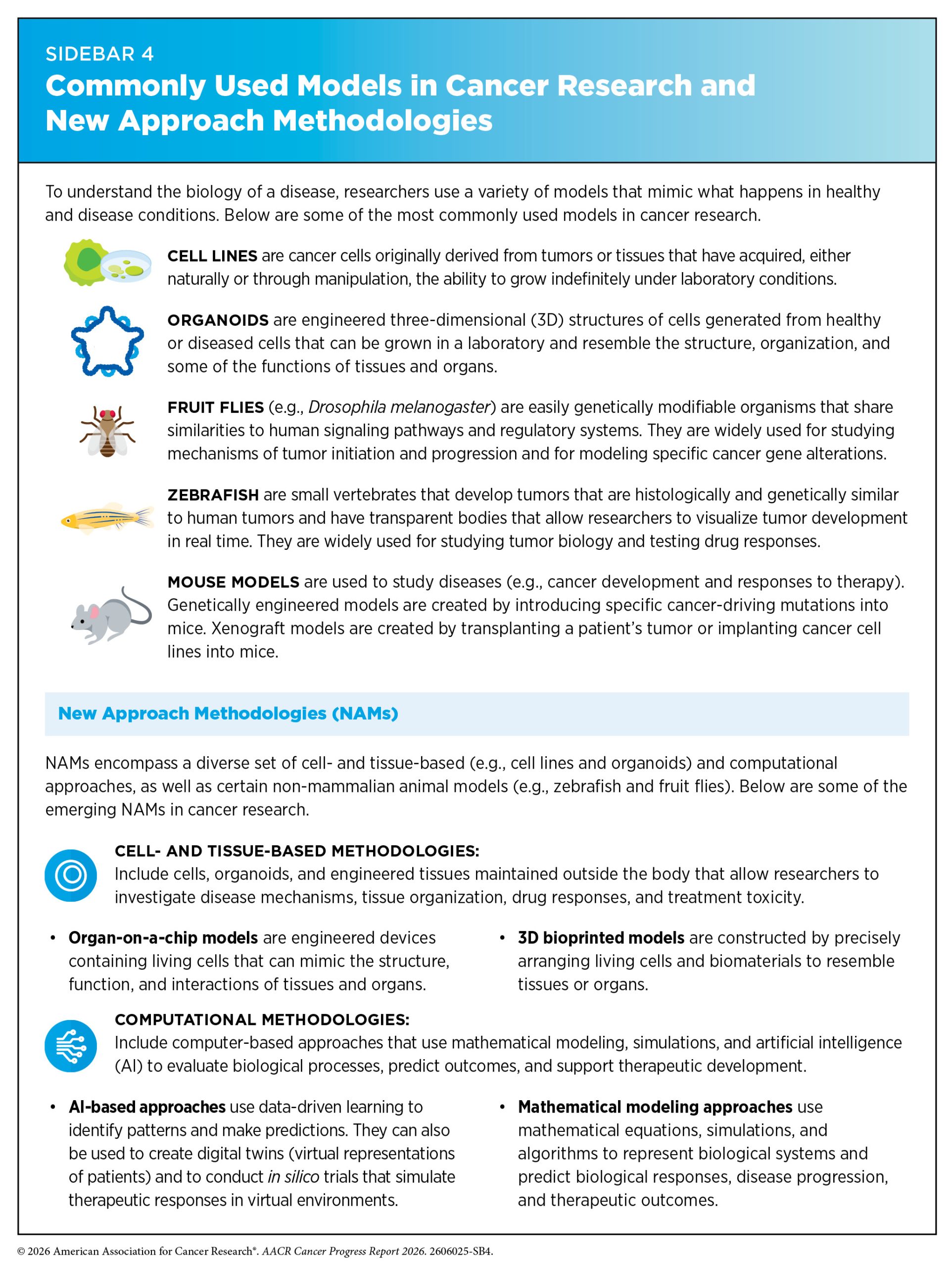
Researchers investigate cancer biology using a wide array of experimental models that mimic healthy and disease conditions (see Sidebar 4). Studies using model organisms have uncovered fundamental biological processes that have shaped our understanding of cancer development. For decades, cancer research has relied heavily on animal models to study tumor development and evaluate potential therapies. More recently, advances in tissue engineering, stem cell biology, biomaterials fabrication, and computational science have led to the emergence of new approach methodologies (NAMs), which can include non-mammalian animals, organoids, organ-on-a-chip systems, artificial intelligence (AI)–based applications, and other approaches that can complement and reduce reliance on traditional animal models. Reflecting the growing potential of these approaches, NIH and the United States (US) Food and Drug Administration (FDA) have recently expanded efforts to broaden the use of NAMs in biomedical research (see Rapidly Delivering Safe and Effective Therapies to Patients). Recent efforts include the launch of NIH’s Office of Research Innovation, Validation, and Application, which coordinates NIH-wide efforts to advance NAMs, and the Complement Animal Research in Experimentation (Complement-ARIE) program, which supports the development, validation, standardization, and adoption of these approaches. In March 2026, NIH announced more than $150 million in funding to develop and scale NAMs, marking the first awards under the Complement-ARIE program (93)National Institutes of Health. NIH invests $150 million in human-based research to reduce use of animal models. Accessed: June 30, 2026.. At the same time, FDA has announced plans to reduce reliance on animal testing for the development of certain drugs and issued guidance on incorporating NAMs into drug development and regulatory decision-making (94)US Food and Drug Administration. FDA Issues Draft Guidance to Cut Unnecessary Animal Testing for Cancer Drugs. Accessed: June 30, 2026.. Ongoing efforts are focused on defining the contexts in which NAMs can complement existing research models while addressing remaining barriers to their effective integration into biomedical research (91)Hanahan D (2026) Cell, 189: 2254.(95)Harrison C (2025) Lab Anim (NY), 54: 250.(96)Lloyd KCK (2025) Nature, 646: 802..
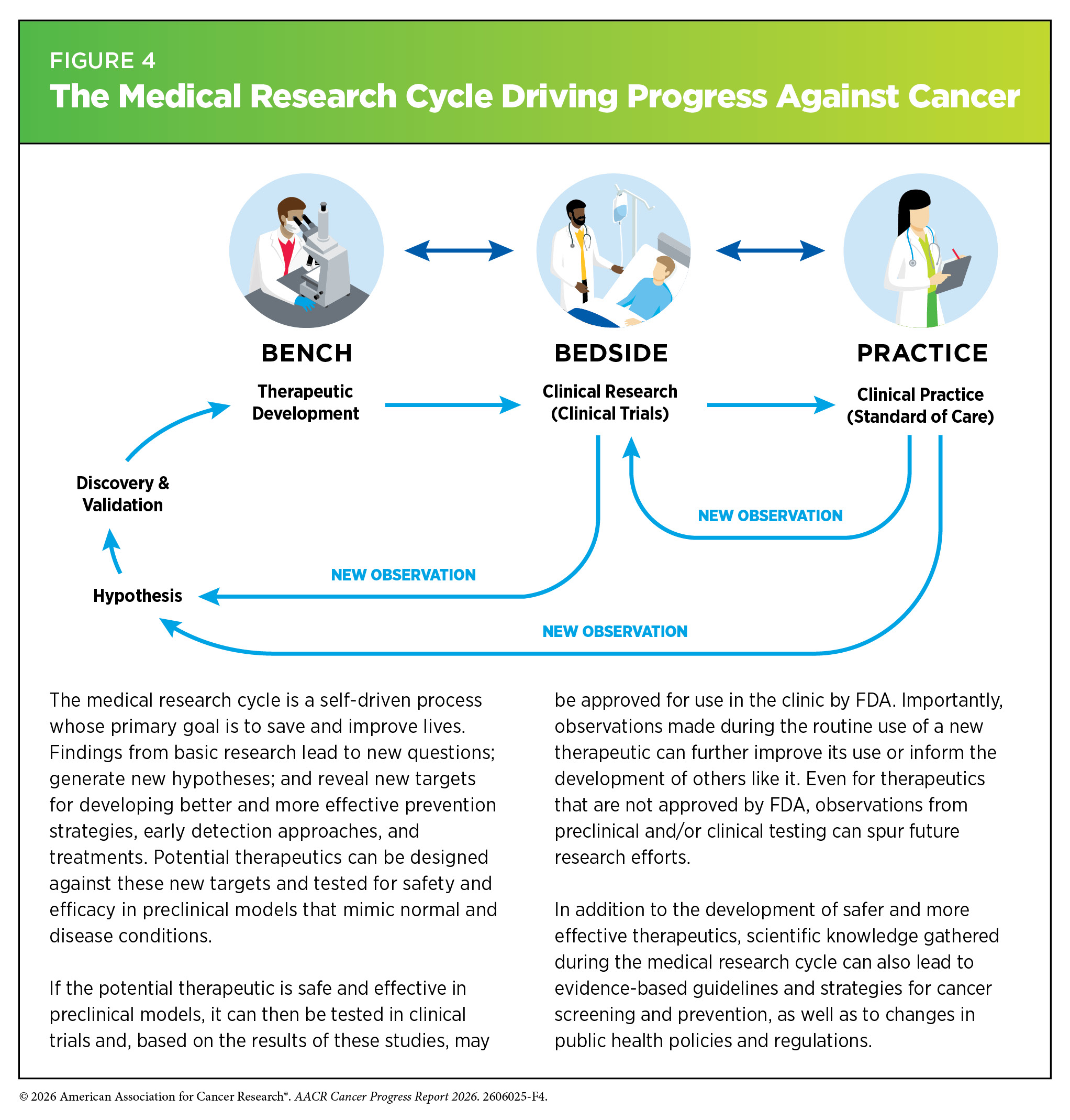
Findings and hypotheses stemming from basic research are fundamental to understanding what triggers cancer development, how cancer evades the body’s defenses, and how cancer spreads within the body. Basic research plays a central role in the medical research cycle (see Figure 4).
Collectively, knowledge gained from medical research has advanced cancer risk reduction, enabled innovations in early detection and diagnosis, and improved personalized treatments. These advances are contributing to progress against cancer that is saving lives and improving health outcomes for countless patients.
Basic Research Decoding Cancer’s Complexities
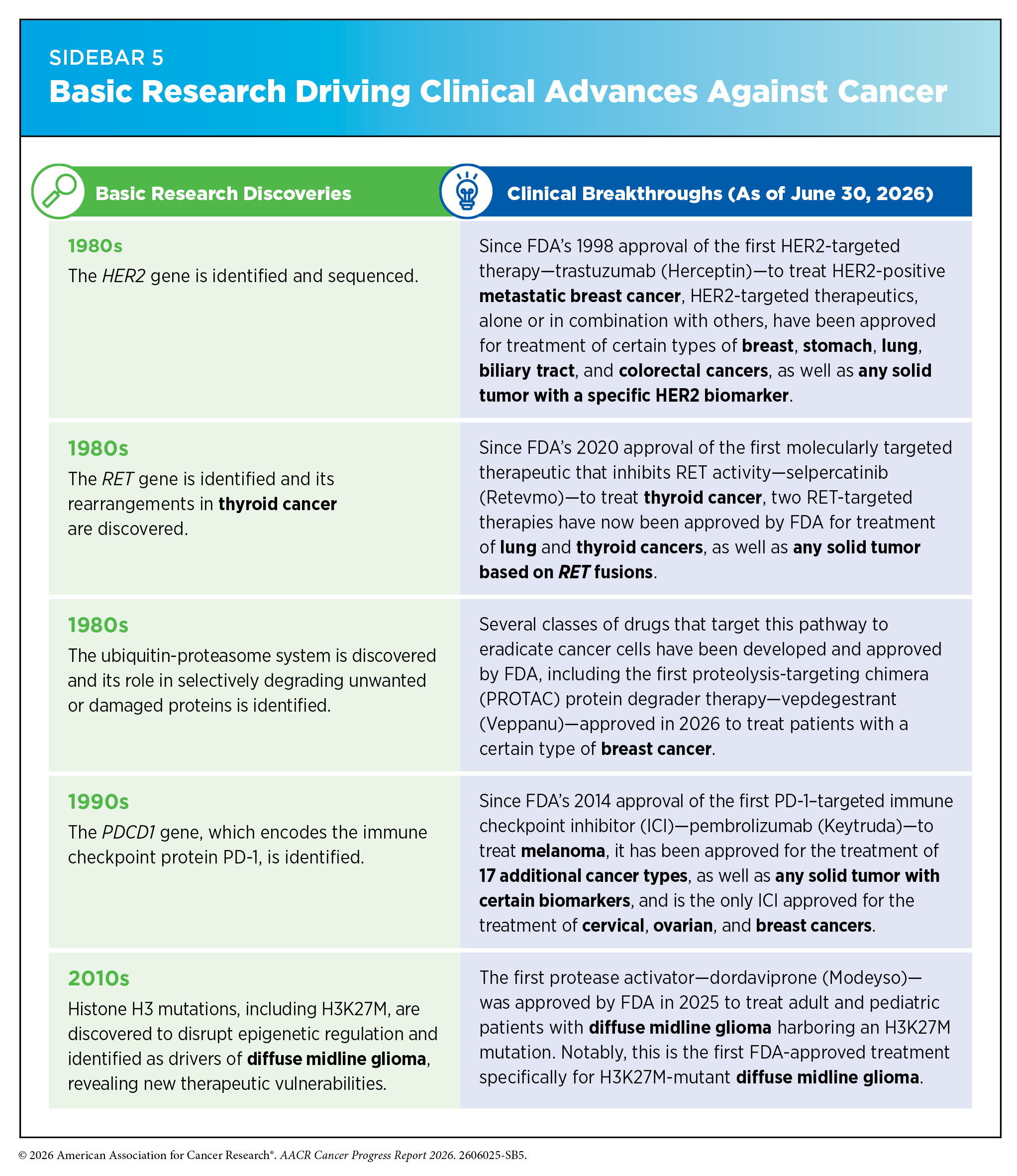
Decades of basic research, and discoveries stemming from it, have provided the foundation for progress against cancer (see Sidebar 5). More than 50 years ago, studies of cancer-driving viruses fundamentally changed our understanding of cancer. Researchers investigating how certain viruses caused tumors discovered that the cancer-driving genes carried by these viruses originated from normal genes already present in healthy cells. This landmark discovery established the concept that cancer can arise when changes in the body’s own genes transform normal genes into cancer-driving oncogenes. Building on these discoveries, researchers identified the first human oncogenes, including three closely related RAS genes (HRAS, KRAS, and NRAS), revealing that mutations in these genes can drive cancer development.
One notable example of how these fundamental discoveries have transformed cancer care is the development of therapies targeting Kirsten rat sarcoma viral oncogene homolog (KRAS), a protein encoded by the KRAS gene (see Figure 5). The KRAS gene is altered in roughly 30 percent of human cancers, including many pancreatic, lung, and colorectal cancers. Once thought to be an “undruggable” target because of its complex structure and function, KRAS has now become one of the most rapidly advancing areas of precision oncology, demonstrating how decades of basic research can lead to lifesaving therapies.
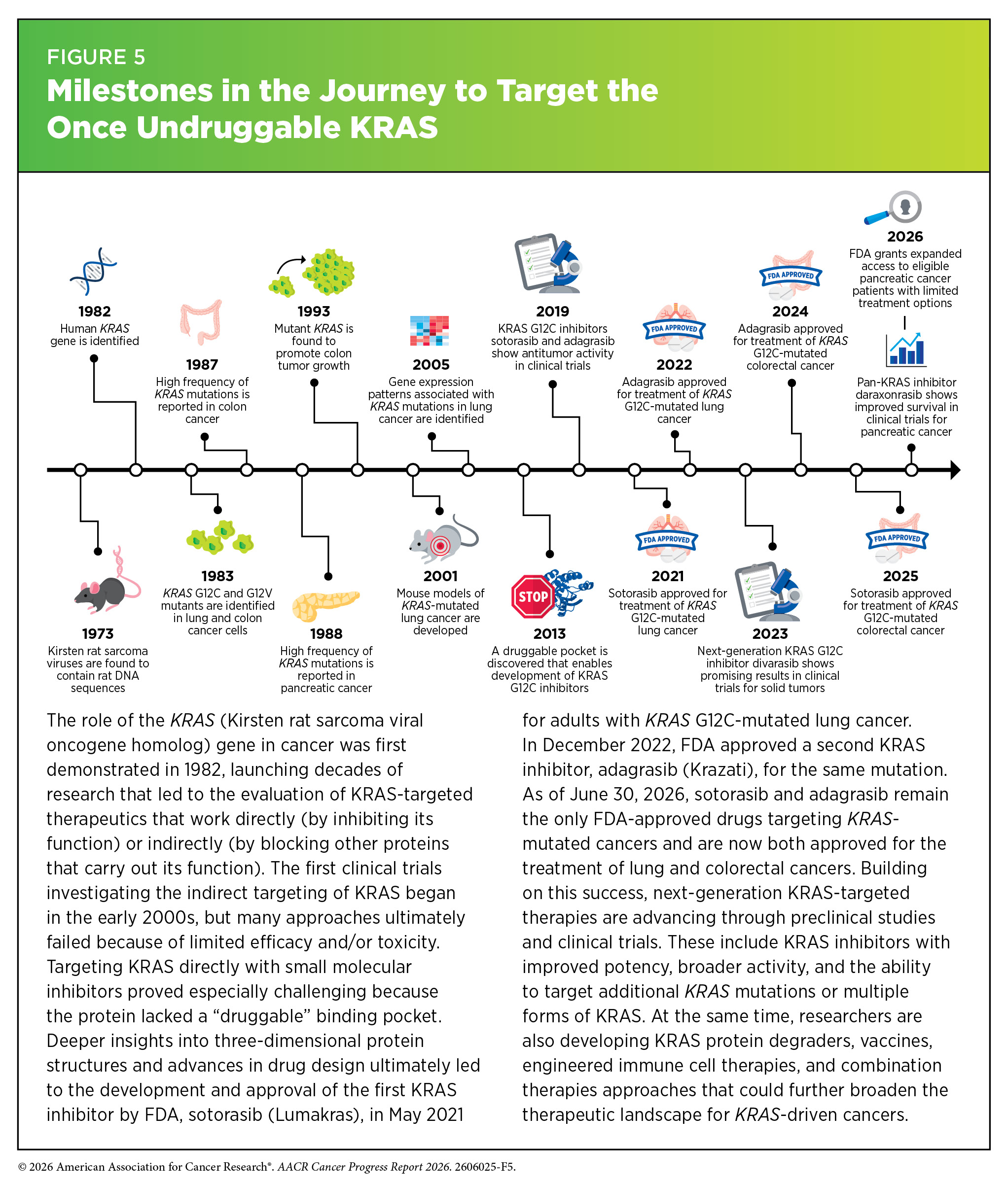
In 1982, researchers found that mutations in the gene could drive cancer development. Over the following decades, basic research revealed that the KRAS protein functions as a molecular switch that cycles between active and inactive states to regulate cell growth, survival, and other essential cellular processes. Mutations can cause the protein to be locked in the “on” position, allowing cells to grow uncontrollably. Although these discoveries established that KRAS mutations could drive cancer, the KRAS protein lacked binding pockets for therapeutic molecules to target, presenting a major challenge for drug development.
Progress toward overcoming this challenge was eventually made possible through a series of fundamental research breakthroughs. Advances in understanding the three-dimensional (3D) structure of proteins uncovered a hidden pocket on a mutant form of KRAS, called KRAS G12C, and new chemistry tools helped researchers design drugs that bind to that pocket and inhibit the protein’s cancer-driving activity.
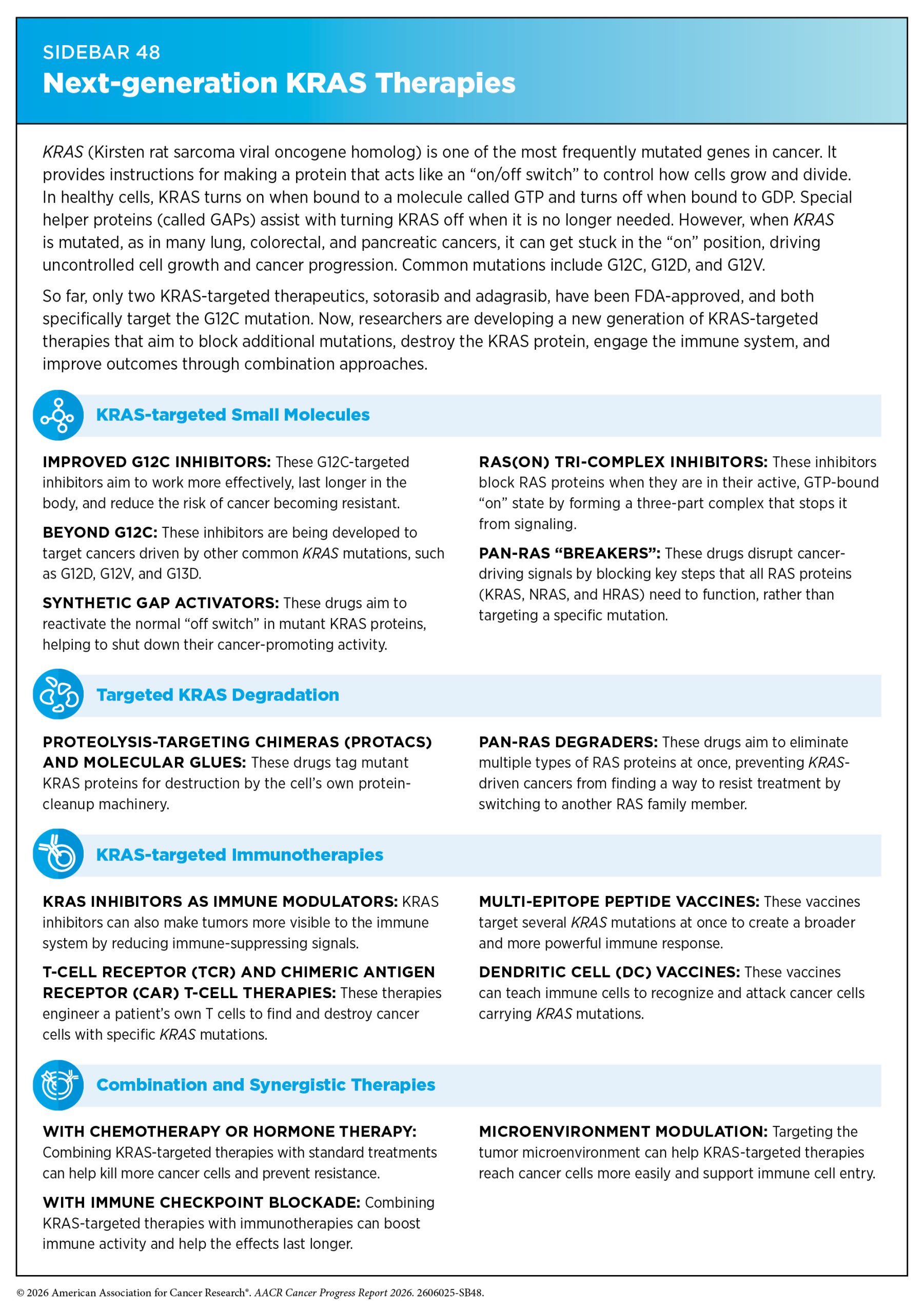
This breakthrough laid the foundation for the development of sotorasib (Lumakras), the first FDA-approved KRAS inhibitor for adults with KRAS G12C-mutated non–small cell lung cancer in 2021, nearly 40 years after the role of the KRAS gene in cancer was discovered. This milestone was quickly followed by the 2022 FDA approval of adagrasib (Krazati), a second KRAS inhibitor targeting the same mutation (see Sidebar 48). As of June 30, 2026, sotorasib and adagrasib remain the only FDA-approved drugs targeting KRAS-mutated cancers, now approved for both lung and colorectal cancers, with ongoing research focused on enhancing their effectiveness and expanding their use across additional cancer types.
However, the impact of these foundational basic research discoveries extends far beyond these first-generation KRAS inhibitors. Next-generation KRAS inhibitors are advancing through clinical trials with improved potency, broader activity, and the ability to target additional KRAS mutations (e.g., the KRAS G12D mutation common in pancreatic cancer) or work more broadly against multiple forms of KRAS—known as pan-KRAS inhibitors (97)Riedl JM, et al. (2026) Cancer Cell, 44: 471.. As one example, a recent phase III clinical trial showed that the pan-RAS inhibitor daraxonrasib significantly improved outcomes for patients with metastatic pancreatic cancer. Compared to standard chemotherapy, treatment with daraxonrasib doubled both median overall survival (13.2 vs. 6.7 months) and progression-free survival (7.3 vs. 3.5 months) (98)O’Reilly EM, et al. (2026) N Engl J Med..
Insights gained through decades of research are also driving the development of entirely new therapeutic approaches beyond KRAS inhibitors (99)Choucair K, et al. (2025) Signal Transduct Target Ther, 10: 385.. These include protein degraders designed to eliminate KRAS proteins from cancer cells, vaccines that train the immune system to recognize KRAS-mutant tumors, engineered immune cells that selectively attack cancer cells carrying KRAS mutations, and combination strategies that simultaneously target multiple pathways involved in KRAS-driven cancers (100)Park W, et al. (2026) N Engl J Med, 394: 1409.(101)Wainberg ZA, et al. (2025) Nat Med, 31: 3648.(102)Benton A, et al. (2025) Cancer Cell, 43: 1365.(103)Modest DP, et al. (2025) Lancet Oncol, 26: 1240.(104)Huff AL, et al. (2026) Nat Commun, 17: 1538.. Together, these approaches have transformed KRAS from a single therapeutic target into one of the most active areas of cancer drug development, with the potential to improve outcomes for patients with a wide range of cancers.
Translating Knowledge to Understand the Biology of Cancer
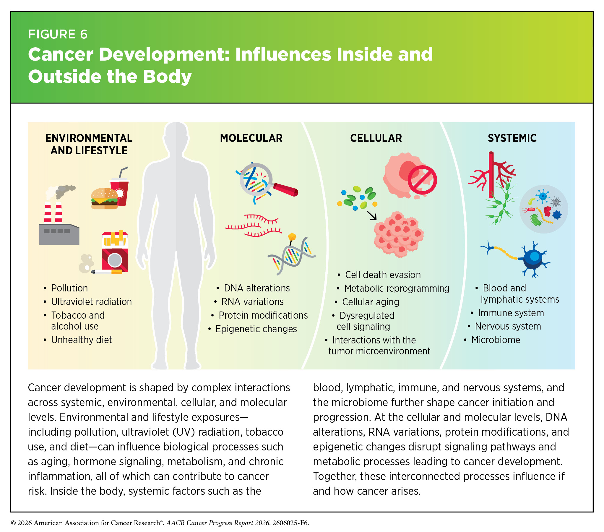
Cancer development is a complex and multistep process shaped by interactions among influences outside the body, throughout the body, within tissues, and inside individual cells (see Figure 6). Influences outside the body, including social drivers of health, behavioral risk factors (e.g., smoking and alcohol consumption), and environmental exposures (e.g., air pollution and environmental carcinogens), affect biological processes throughout the body and can contribute to the molecular and cellular changes that drive cancer initiation and progression. Factors such as aging, obesity, and chronic inflammation can also alter the surrounding tissue environment, further facilitating conditions that favor the growth and survival of cancer cells (105)De Dominici M, et al. (2026) Cancer Discov, 16: 16..
As cancer progresses, cells accumulate additional changes and evolve in ways that allow them to grow more aggressively and evade the immune system. Some of these changes allow cancer cells to switch between different cellular states in response to signals and changing conditions in their environment, a process known as cellular plasticity. As these changes accumulate, cancer cells become increasingly diverse.
This continuous evolution, along with cellular plasticity, leads to significant molecular and cellular differences among cells within a single tumor, a phenomenon known as tumor heterogeneity. As one example, researchers recently identified a rare population of highly adaptable cancer cells in lung tumors that can generate diverse cell states and drive tumor growth, progression, and therapy resistance (106)Chan JE, et al. (2026) Nature, 651: 231.. Tumor heterogeneity also describes the differences that can exist between tumors arising in the same tissue type across different individuals, as well as among multiple tumors within an individual patient when the cancer has spread.
Understanding the cellular and molecular factors inside and outside the cell that influence cancer initiation and progression is key to developing more effective strategies for prevention and treatment.
Influences Inside the Cell
Cells store their genetic information in DNA, a double-helical molecule made of four chemical building blocks called bases: adenine (A), thymine (T), cytosine (C), and guanine (G). The complete set of a person’s DNA is called the genome.
In human cells, DNA is wrapped around proteins called histones to form chromatin, which is further compacted into chromosomes. Most human cells contain 46 chromosomes. Each chromosome contains hundreds to thousands of genes, which are segments of DNA that carry the instructions for making proteins. Epigenetic modifications—chemical changes to DNA and histones—help regulate which genes are turned on or off, allowing cells with the same genome to develop into different cell types and perform specialized functions.
To make proteins, cells first copy the instructions encoded in DNA into a molecule called messenger RNA (mRNA) through a process known as transcription. The mRNA is then used as a template to produce proteins, which carry out most of the essential functions of cells. When the genetic instructions or the processes that read and interpret them are altered, the tightly regulated programs that control normal cell growth and function can break down, leading to cancer.
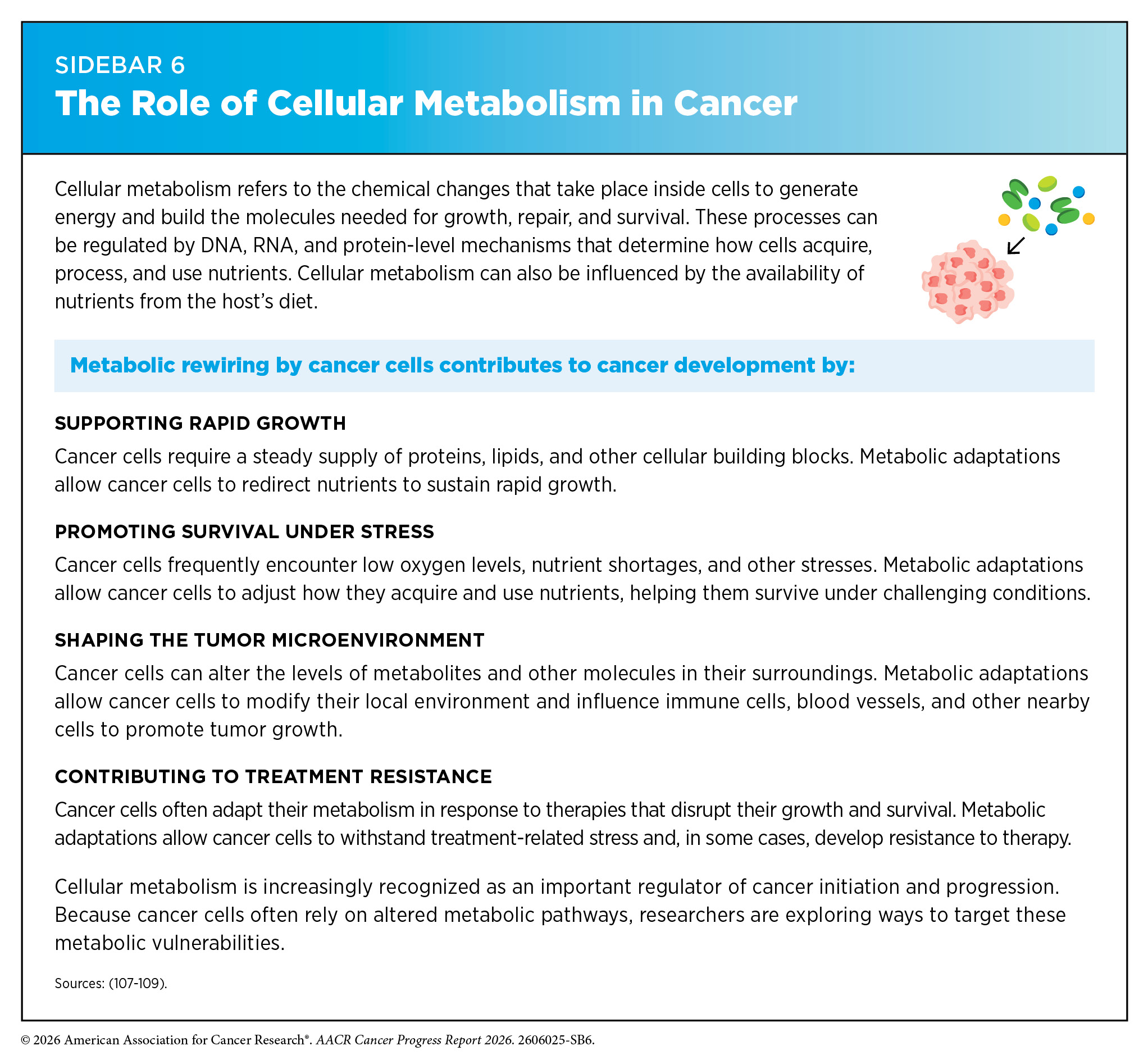
While genetic alterations are often permanent changes to the DNA sequence, many changes involving RNA, proteins, and epigenetic regulation, are more dynamic and can rapidly shift in response to internal and external influences. These molecular changes can also alter metabolic pathways, which determine how cells generate energy, use nutrients, and respond to stress. This metabolic rewiring is increasingly recognized as an important driver of cancer development (see Sidebar 6). The following sections explore the types of changes within the cell that can lead to cancer development.
Genetic Alterations
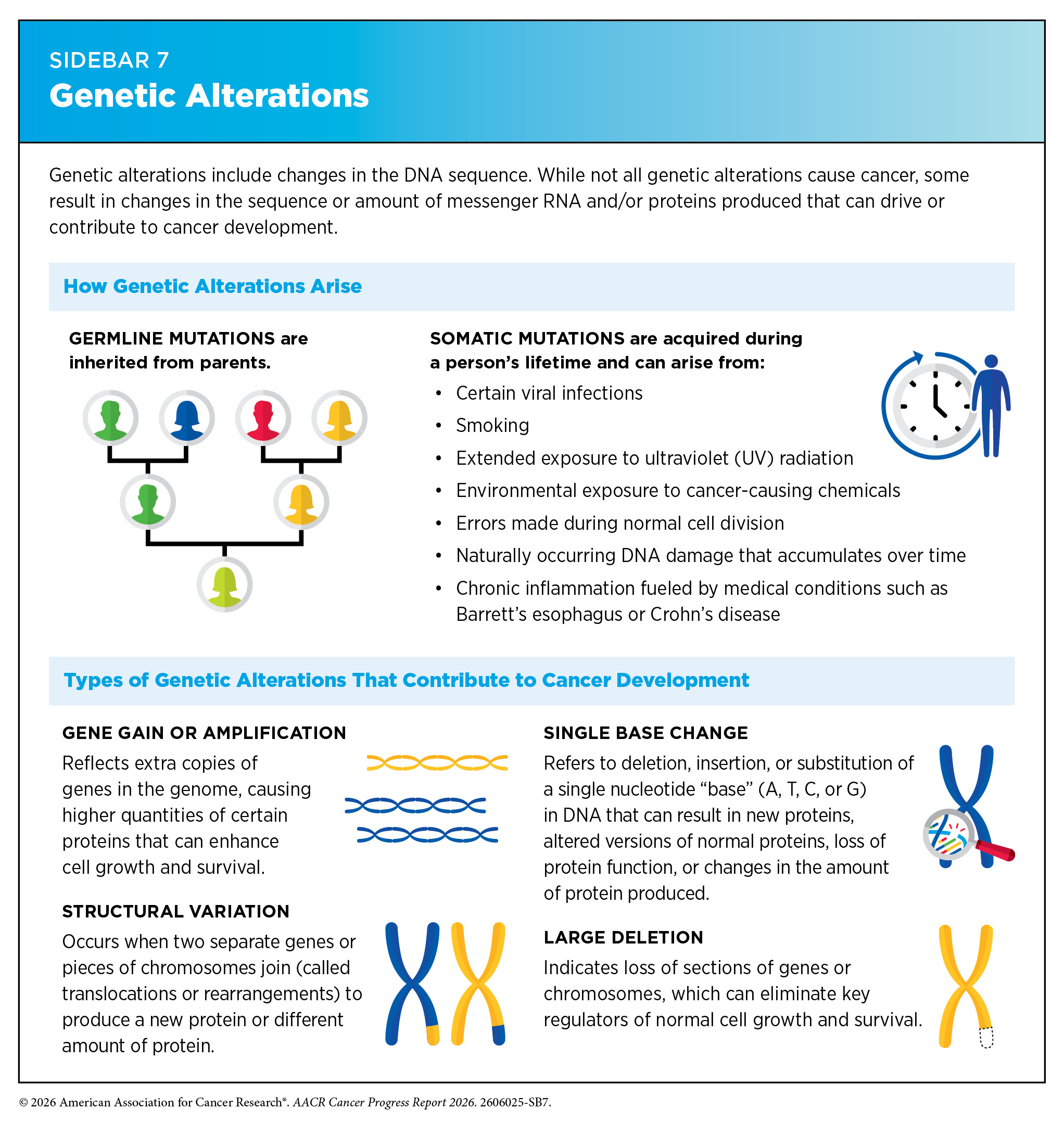
Genetic alterations, also called mutations or variants, include changes in the DNA sequence and can change the sequence or amount of the resulting mRNA and protein, thus disrupting or modifying normal protein function and contributing to cancer development. Genetic alterations can be inherited from parents (referred to as germline mutations) or acquired throughout a person’s lifetime (referred to as somatic mutations) (see Sidebar 7). Whether inherited or acquired, genetic mutations can disrupt normal cellular functions.
Germline mutations play a critical role in determining the inherited risk of developing cancer. About 10 percent of cancer cases are caused by germline mutations. These mutations occur in the body’s reproductive cells (egg or sperm) that are passed on from parents to children and can increase their risk of developing cancer; however, not all germline mutations contribute to cancer development.
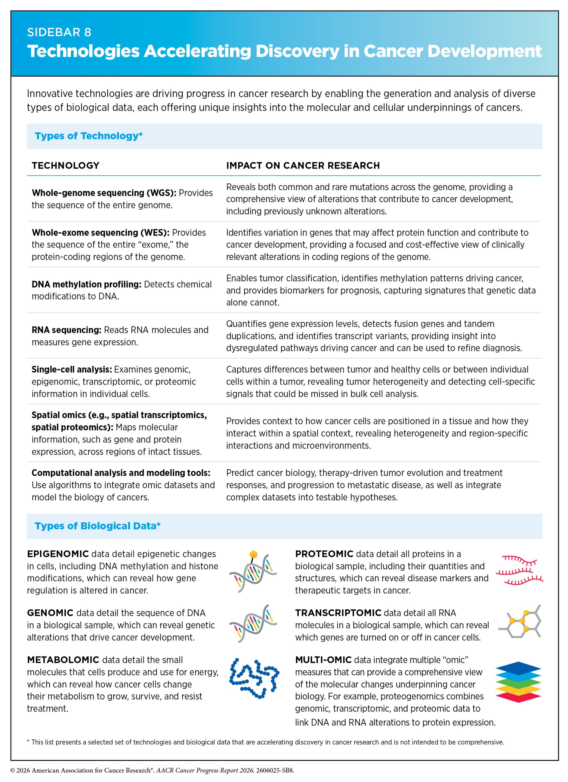
Emerging technologies are expanding our ability to detect a wide spectrum of genetic alterations (see Sidebar 8). Advances in DNA sequencing have enabled a better understanding of germline mutations that are associated with a person’s risk of developing cancer. A recent study analyzing sequences of the protein-coding regions of the genome from more than 820,000 individuals identified inherited variants associated with breast cancer susceptibility, expanding the number of confirmed breast cancer predisposition genes (111)Collister JA, et al. (2025) NPJ Breast Cancer, 11: 111.. These findings may improve our understanding of hereditary breast cancer risk and offer new opportunities to inform risk prediction, prevention, and early detection strategies.
Inherited genetic variants can also influence how acquired mutations evolve over time. Clonal hematopoiesis is a condition in which blood stem cells acquire genetic mutations and give rise to genetically distinct populations of blood cells, and it can increase the risk of developing blood cancers. Research has identified inherited variants that increase susceptibility to clonal hematopoiesis, shape which mutated cells expand, and influence the likelihood that these cells progress to blood cancers (112)Liu J, et al. (2025) Nat Genet, 57: 1872.(113)Weinstock JS, et al. (2025) Nat Commun, 16: 9194..
However, inherited genetic variation does not always increase risk. A recent study identified an inherited variant that reduced the likelihood of clonal hematopoiesis. Researchers found that this variant limited the expansion of mutated blood cells, reducing the risk of clonal hematopoiesis and certain blood cancers. These findings highlight how inherited genetic variation can influence cancer risk not only by increasing susceptibility but also by promoting resilience to cancer-associated cellular changes (114)Agarwal G, et al. (2026) Science, 391: 52..
Large-scale genome-wide association studies—which can analyze millions of genetic variations associated with disease risk—are identifying cancer risk variants at an unprecedented scale. As one example, in the largest genome-wide association study of high-grade serous ovarian cancer to date, researchers analyzed over 22 million genetic variants from nearly 400,000 women and identified seven previously unknown genomic regions associated with disease risk, improving models used to predict inherited susceptibility (115)Barnes DR, et al. (2025) NPJ Genom Med, 10: 73..
Somatic, or acquired, mutations arise during an individual’s lifetime as a result of naturally occurring DNA damage, errors during normal cell division, or in response to environmental exposures, lifestyle factors, or chronic health conditions. These mutations contribute to the unique genetic landscape of individual tumors, with distinct patterns reflecting potential causes of cancer, tissue of origin, and evolution over time.
Somatic mutations accumulate throughout normal tissues over the course of life. Recent advances in highly sensitive DNA sequencing technologies have allowed researchers to identify cancer-associated driver mutations and clonal expansions in normal oral and bladder tissue that would not have been detectable using conventional sequencing approaches (116)Calvet F, et al. (2025) Nature, 647: 436.(117)Lawson ARJ, et al. (2025) Nature, 647: 411.. While clonal hematopoiesis is a well-recognized example of how aging can reshape blood cell populations, similar processes are increasingly being identified in solid tissues. For example, researchers recently found that otherwise normal pancreatic tissue may harbor hundreds of pancreatic intraepithelial neoplasias (PanINs)—the most common precursors to pancreatic cancer. They found that nearly all PanINs contained cancer-associated somatic mutations and the adult pancreas can harbor hundreds of these lesions. Because pancreatic cancer remains relatively uncommon despite this high burden of precursor lesions, they suggest only a small fraction of PanINs progress to invasive cancer (118)Braxton AM, et al. (2024) Nature, 629: 679.. Together, these findings suggest that genetically altered cells and precursor lesions may be far more common than previously thought, with important implications for cancer risk assessment, early detection, and prevention efforts.
Recent research has emphasized the importance of understanding how somatic mutation patterns can vary by age, as these differences may influence cancer initiation, progression, and response to treatment. The incidence of early-onset cancers, defined as cancers diagnosed in individuals ages 18 to 49, is growing globally (see The Growing Population Burden of Cancer). A large international study of more than 17,000 colorectal cancer (CRC) patients found that tumors in younger adults often carry different genetic alterations than those diagnosed later in life. These findings support the need for age-informed molecular profiling that may inform future approaches to cancer diagnosis and treatment (119)Li J, et al. (2025) Lancet Oncol, 26: 1055..
In addition to germline and somatic DNA sequence mutations, structural genomic alterations—including chromosomal rearrangements and nontraditional DNA structures—can also influence cancer development. For example, chromosomal rearrangements can generate cancer-causing gene fusions, in which parts of two genes become joined to create a hybrid gene that produces an abnormal fusion protein capable of driving cancer development.
Extrachromosomal DNA (ecDNA)—circular DNA fragments that exist outside chromosomes—is an example of a nontraditional DNA structure that has emerged as a key contributor to tumor growth, treatment resistance, and poor clinical outcomes across cancer types. By carrying genes that regulate cell growth and survival, ecDNA acts as a potent driver of cancer progression. Unlike chromosomal DNA, ecDNA does not follow the normal rules of inheritance during cell division, enabling rapid amplification of cancer-promoting genes and allowing cancer cells to adapt more easily to treatments and other stresses in the tumor microenvironment.
Cutting-edge technologies and computational analysis tools have revealed that ecDNA is present in approximately 17 percent of human cancers, although the prevalence varies considerably across cancer types. For example, ecDNAs have been detected in 30 percent of pediatric neuroblastomas and 49 percent of adult glioblastomas, but in only 3 percent of kidney cancers (121)Wong IT, et al. (2026) Cell, 189: 2307.. Just in the past year, notable progress has been made in ecDNA detection (122)Chowdhury B, et al. (2025) Nat Commun, 17: 894. and our understanding of how ecDNA contributes to cancer development, including its role in driving oncogene overexpression (123)Guan R, et al. (2026) Oncogene, 45: 1100.(124)Montuori G, et al. (2025) Cancer Discov, 15: 2054., increasing tumor heterogeneity (125)Krupina K, et al. (2025) Science, 390: 1156.(126)Noorani I, et al. (2025) Cancer Discov, 15: 2078., promoting immune evasion (127)Zhang J, et al. (2025) Clin Cancer Res, 31: 4529.), and enabling resistance to therapy (124)Montuori G, et al. (2025) Cancer Discov, 15: 2054..
New research has revealed several potential therapeutic vulnerabilities in ecDNA-driven cancers. These include dependencies on pathways that help cells resolve conflicts between the cell’s DNA copying and RNA producing machineries, mechanisms that maintain ecDNA structure and inheritance during cell division, and molecular interactions that enable ecDNA to drive high levels of oncogene expression (128)Wong IT, et al. (2026) Nat Rev Drug Discov, 25: 374.(129)Sankar V, et al. (2026) Nature, 649: 152.(130)Taghbalout A, et al. (2025) Cancer Cell, 43: 2191.(131)Kraft K, et al. (2025) Nat Cell Biol, 27: 1914.. Reflecting the importance of ecDNA, the Cancer Grand Challenges, a global multidisciplinary research initiative, is funding research into how ecDNA forms, functions, and can be targeted in cancer (see Technological Innovations and Collaborative Science).
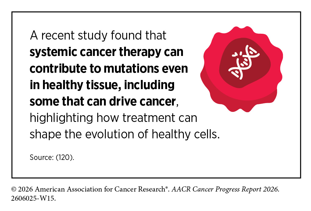
Researchers are building on the understanding of genetic alterations that drive cancer development to inform new strategies for cancer prevention, diagnosis, and treatment. Over the past two decades, FDA has approved numerous targeted therapies designed to inhibit the effects of specific genetic alterations found in cancer cells (132)National Cancer Institute. Targeted Therapy Drug List by Cancer Type. Accessed: June 30, 2026..
At the same time, genetic tests that detect germline mutations are helping individuals understand their inherited cancer risk, guiding clinical decisions, and supporting proactive health management for both patients and their families (see Advances in Genomics-based and Risk-stratified Precision Approaches).
RNA Variations
Gene expression is the process by which a cell reads the instructions in a gene to produce mRNA. Cells can regulate this process to increase or decrease mRNA production as needed to respond to internal and external signals. In cancer, these regulatory processes often go awry.
Most genes are made up of alternating DNA segments known as exons and introns. Exons contain the instructions needed to make proteins, whereas introns do not. When a gene is transcribed into mRNA, the resulting transcript includes both exons and introns. Through a “cut and paste” process called RNA splicing, introns are removed and exons are joined together to produce an mRNA molecule that can then be translated into a functional protein. Cells also use a mechanism called alternative splicing to produce mRNAs with different combinations of exons from a single gene, generating multiple protein variants with distinct cellular functions.
If errors occur during RNA splicing, faulty mRNAs can produce dysfunctional proteins. In a recent study, aberrant splicing driven by loss of the MEN1 gene was found to play a critical role in driving cancer growth and metastasis in lung cancer (133)Pan T, et al. (2026) Cancer Res, 86: 146.. Researchers are exploring the potential of targeting RNA splicing mechanisms as innovative therapeutic strategies to restore normal splicing patterns or selectively eliminate aberrant splice variants that are associated with cancer (134)Kral AJ, et al. (2026) Mol Cell, 86: 60.(135)Wagner V, et al. (2026) Nat Commun, 17..
In addition to mRNA, cells also produce non-coding RNAs (ncRNAs)—RNA molecules that do not encode proteins but play critical roles in regulating gene expression. These include microRNAs (miRNAs), long non-coding RNAs (lncRNAs), and circular RNAs (circRNAs), each of which can drive normal cellular processes but also influence how tumors grow, interact with their environment, and resist treatment (136)Hashemi M, et al. (2025) Cancer Cell Int, 25: 350.(137)Bitaraf A, et al. (2025) Mol Cancer, 24: 245.(138)Xue Z, et al. (2026) Mol Cancer Res, 24: 283..
While some RNA variations arise from changes in how RNA molecules are processed or regulated, such as aberrant splicing or dysregulated ncRNAs, others reflect the downstream effects of genetic alterations in DNA. Understanding how tumors change at the RNA level therefore provides vital insights into how cancer develops, evolves, and spreads. Advances in transcriptomic and RNA sequencing technologies are being used to identify and understand RNA-level changes by mapping how genes are expressed within individual tumor cells and across different regions of a tumor (see Sidebar 8).
A growing proportion of lung cancer cases are being diagnosed in individuals who have never smoked (see The Growing Population Burden of Cancer) (53)Caswell DR, et al. (2026) Trends in Cancer, 12: 310.. Although previous studies have identified many genetic alterations associated with these cancers (139)Díaz-Gay M, et al. (2025) Nature, 644: 133., less is known about how these tumors differ at the transcriptomic level. In a recent study, researchers conducted a large-scale transcriptomic analysis of tumors from more than 680 never-smoker lung cancer patients and identified three distinct transcriptomic subtypes with unique gene expression patterns and cellular characteristics associated with differences in clinical outcomes (140)Zhao W, et al. (2026) Cancer Discov, 16: 460.. These findings may help guide the development of more personalized approaches to diagnosis and treatment in this patient population.
Additionally, researchers are using RNA sequencing to reveal the functional consequences of genetic alterations at the RNA level, helping to advance precision medicine. In a study of more than 2,300 children and adults with a wide range of solid tumors and blood cancers, targeted RNA sequencing identified clinically relevant RNA alterations that refined diagnoses and informed treatment decisions in 87 percent of cases (141)Siddaway R, et al. (2025) Nat Med, 31: 3524..
As researchers continue to uncover how RNA variations contribute to cancer development, these discoveries are opening new therapeutic opportunities. Advances in RNA biology are enabling the development of RNA-based medicines that can regulate gene expression, produce therapeutic proteins, and stimulate antitumor immune responses (see RNA Medicines).
Protein Modifications
Proteins are vital for normal cellular functions. The human genome contains approximately 20,000 protein-coding genes, and the full set of proteins expressed by the cell is known as the proteome. Proteomics is the study of the proteome, which can provide new insights into cancer biology and reveal molecular features that may help guide diagnosis and treatment.
Researchers recently expanded on earlier proteomic studies, which mostly focused on individual cancer types, by generating a pan-cancer proteomic atlas comprising more than 9,600 proteins from nearly 1,000 tumors representing 22 cancer types. This comprehensive resource revealed both shared and cancer-specific molecular features and identified potential biomarkers and therapeutic targets (142)Knol JC, et al. (2025) Cancer Cell, 43: 1328..
Proteogenomic research integrates protein data with genomic, transcriptomic, and other molecular data to provide a more comprehensive view of the molecular changes driving cancer than any single type of analysis alone (see Sidebar 8). Researchers recently analyzed nearly 400 tumors from patients with medulloblastoma—a fast-growing brain cancer common in children and young adults—revealing previously unrecognized tumor subtypes based on their protein and metabolic features, uncovering biological insights not apparent from genomic and transcriptomic analyses alone, and identifying a therapeutically targetable metabolic vulnerability (143)Bernardi F, et al. (2026) Cancer Cell, 44: 383..
The functions of many proteins are regulated by posttranslational modifications (PTMs), which are reversible chemical changes that modulate protein function, localization, and stability. Advances in AI are accelerating research across the cancer continuum (see Artificial Intelligence), including in cancer proteomic research. Researchers have recently developed an AI model, trained on a dataset containing nearly 400,000 experimentally validated PTM sites, to predict how genetic variants alter PTMs (144)Wen B, et al. (2025) Nat Methods, 22: 1857.. This approach provides new insights into how genetic alterations can disrupt protein regulation and influence disease-associated biological processes, including those involved in cancer development.
Proteins carry out nearly all the cell’s functions and must be located in the right place, in the correct 3D structure, and precisely controlled to function correctly. When proteins are no longer needed or become damaged, cells rely on carefully controlled degradation systems to remove them. Recent research has shown that disruption in the normal protein degradation pathway can contribute to therapeutic resistance. For example, researchers found that impaired degradation of KRAS protein promoted resistance to KRAS-targeted therapies in lung cancer (145)Ivanisevic T, et al. (2025) Nat Commun, 17: 411.. They also identified combination treatment strategies that restore drug sensitivity by blocking signaling pathways activated by impaired KRAS degradation, highlighting potential approaches for overcoming therapeutic resistance.
Researchers are also harnessing the cell’s natural protein degradation machinery as a therapeutic strategy. For example, proteolysis-targeting chimeras (PROTACs) are a new class of therapeutics designed to selectively tag disease-causing proteins for destruction by the cell’s protein degradation system. In May 2026, the first PROTAC therapy, vepdegestrant (Veppanu), was approved by FDA for patients with a certain type of breast cancer (see Advances in Treatment With Molecularly Targeted Therapeutics). This milestone demonstrates how a deeper understanding of protein degradation pathways can lead to new treatment approaches for cancer.
Epigenetic Changes
Epigenetic modifications are chemical changes that regulate how genes are expressed without altering the underlying DNA sequence. The entirety of epigenetic changes within a cell is called the epigenome. Epigenetic changes are shaped by aging, environmental exposures, behavioral and lifestyle factors, and chronic stress. In healthy cells, these mechanisms help to tightly regulate gene expression, but in cancer this regulation is disrupted.
One common type of epigenetic alteration is DNA methylation, in which methyl groups are added to specific regions of the genome to regulate whether nearby genes are turned on or off. Other epigenetic changes, such as chemical modifications of histone proteins and changes in chromatin organization, also influence how genes are expressed and can contribute to cancer development.
In 2021, the World Health Organization published an updated classification of central nervous system (CNS) tumors that recognized DNA methylation profiling as an important tool for classifying and diagnosing these tumors, providing more precise tumor classification than traditional tissue morphology-based analysis and reducing unclear diagnoses (146)International Agency for Research on Cancer. Central Nervous System Tumours. WHO Classification of Tumours, 5th Edition, Volume 6. Accessed: June 30, 2026.. Building on this, researchers recently expanded a widely used DNA methylation–based classification system for CNS tumors by incorporating newly identified tumor subclasses defined by distinct DNA methylation patterns, more than doubling the number of recognized subclasses and improving the identification of rare and previously unrecognized tumor types that can inform clinical decision-making (147)Sill M, et al. (2026) Cancer Cell, 44: 340..
Methylation profiles are also increasingly being coupled with AI to improve diagnostic speed and accuracy. Recently, an AI-assisted framework was developed that uses DNA methylation patterns to classify acute leukemias, a group of aggressive blood cancers that require prompt diagnosis and treatment. The approach identified dozens of distinct leukemia subtypes and accurately classified patients within hours, which could accelerate treatment planning and potentially limit treatment delays (148)Steinicke TL, et al. (2025) Nat Genet, 57: 2456..
Research has also shown that DNA methylation profiles can help predict response and survival in certain patients treated with immune checkpoint inhibitors (ICIs) (see Releasing the Brakes on the Immune System). In a recent study, researchers identified four tumor subtypes of pleural mesothelioma, an aggressive cancer often linked to asbestos exposure (see Limit Exposure to Environmental Risk Factors), with increasing levels of methylation that differed in immune activity and clinical outcomes. They found that tumors with lower levels of DNA methylation showed a more active immune microenvironment and were associated with better responses to ICI therapy and longer survival, whereas highly methylated tumors exhibited features associated with immune suppression and poorer survival (149)Calabrò L, et al. (2026) Nat Genet, 58: 1100..
Epigenetic regulation is also influenced by aging, the immune system, and chronic stress. Researchers often refer to epigenetic clocks as an estimate of a person’s biological age based on their DNA methylation patterns. When an individual’s biological age outpaces their chronological age—known as epigenetic age acceleration—it may signal increased cancer risk. A recent study found that accelerated biological aging, measured by DNA methylation–based epigenetic clocks in blood cells, was associated with an increased risk of developing CRC in postmenopausal women (150)Jung SY, et al. (2025) Aging (Albany NY), 17: 1624.. These findings suggest that epigenetic aging biomarkers could help identify individuals at elevated risk for developing cancer in an aging population.
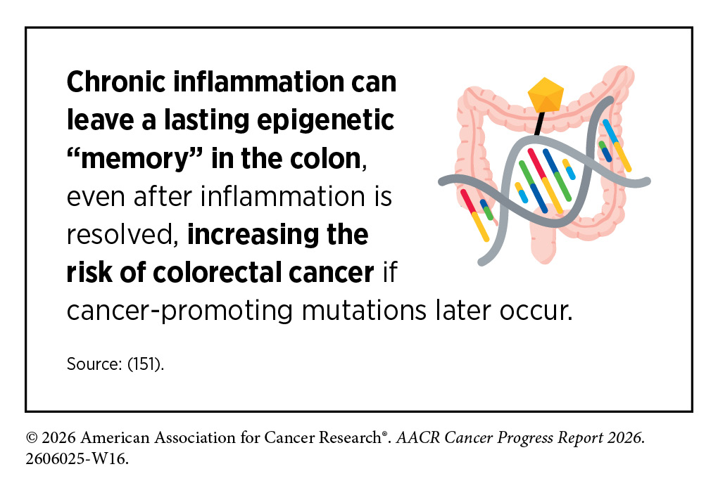
Emerging evidence suggests that epigenetic aging may also play a role in the development of early-onset CRC. Research has found that patients with early-onset CRC exhibit epigenetic changes linked to chronic inflammation and accelerated epigenetic aging, having, on average, an epigenetic age 12 years older when compared to the average-onset CRC patients (152)Jin N, et al. (2025) Cancer Res Commun, 5: 1985..
Research has significantly advanced our understanding of how the epigenome is modified in cancer and how these changes contribute to cancer development. A unique feature of epigenetic changes is that they are dynamic and reversible, making them attractive targets for cancer treatment. Consequently, several anticancer drugs that modify the cancer epigenome have been developed and approved by FDA.
Influences Outside the Cell
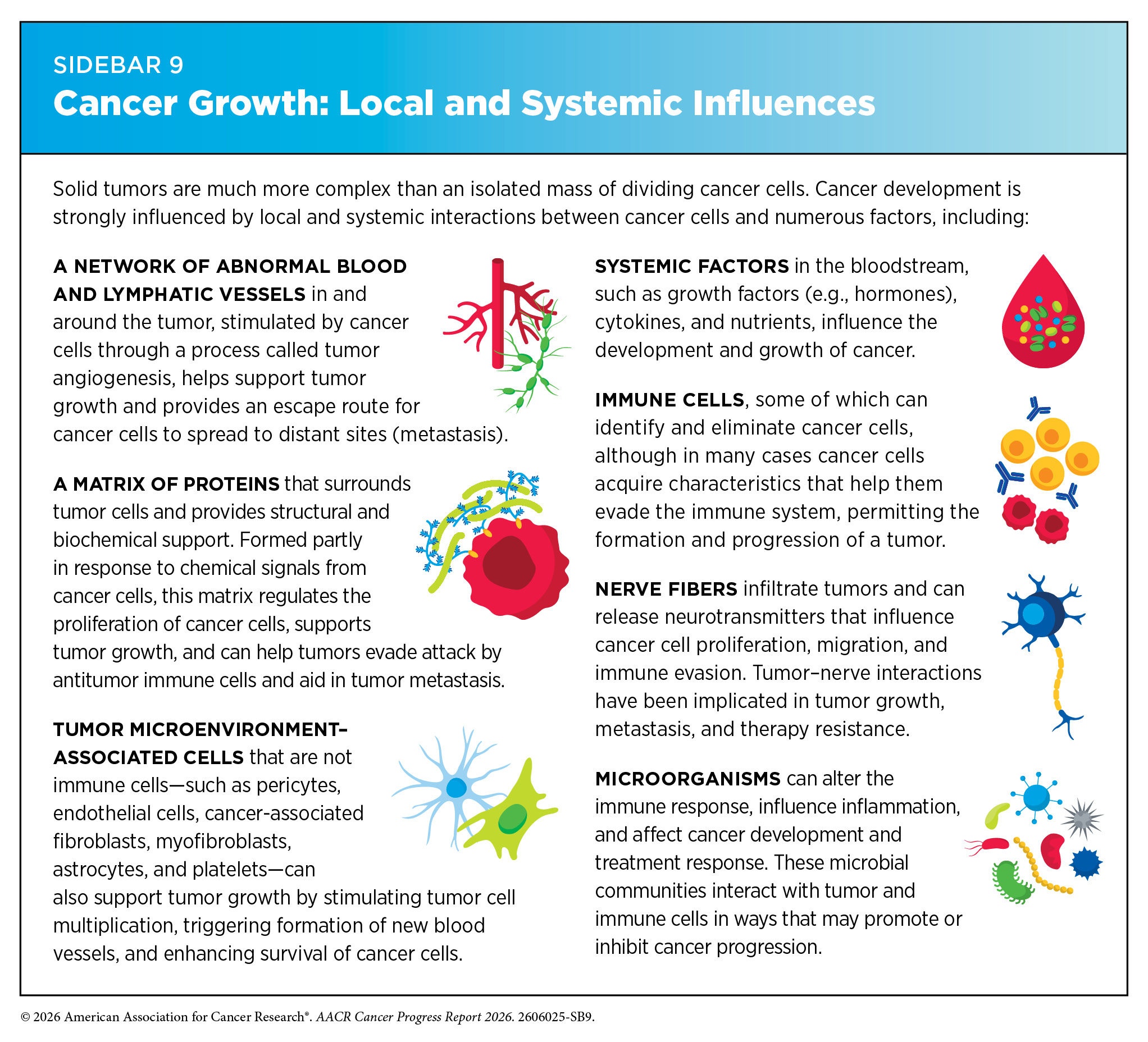
In addition to alterations inside the cell, influences outside the cell contribute to cancer initiation, progression, and response to therapy (see Sidebar 9). These external factors include systemic influences, such as hormones and nutrients in circulation, as well as local tissue environments that interact continuously with tumor cells. A hallmark of cancer is the ability of tumor cells to break away from the primary tissue and travel to other parts of the body (91)Hanahan D (2026) Cell, 189: 2254.. This process—known as metastasis—is enabled by the blood and lymphatic systems, which provide physical routes through which cancer can spread, and the immune system, which can either suppress or support tumor progression, depending on how it is activated or suppressed.
Emerging evidence also shows the nervous system plays an active role in the ability of tumor cells to invade surrounding tissues and communicate with distant organs, influencing tumor growth, invasion, and metastasis. The microbiome—comprising a diverse community of microorganisms residing on and in the body—can influence cancer development by modulating inflammation, immune responses, and systemic metabolism. The tumor microenvironment (TME) encompasses the cellular and structural context in which a tumor exists and shapes how cancer evolves and responds to treatment. The following sections describe the influences outside the cell that can contribute to cancer development.
Blood and Lymphatic Systems
The blood and lymphatic systems are composed of interconnected networks of vessels, circulating cells, signaling molecules, and lymph nodes, as well as several organs, including the spleen, thymus, tonsils, and adenoids. Together, these networks deliver oxygen and nutrients throughout the body, remove cellular waste from tissues, and support the immune system. Additionally, blood and lymphatic vessels provide pathways through which cancer cells can spread from the primary tumor to other parts of the body.
One hallmark of cancer cells is the ability to promote angiogenesis—the formation of new blood vessels—toward and within a tumor, increasing tumor vascularization to help supply the oxygen and nutrients needed to support tumor growth. Unlike normal blood vessels, these newly formed tumor-associated blood vessels are structurally and functionally abnormal. Cancer cells can produce and release high levels of vascular endothelial growth factors (VEGF), a family of signaling proteins that stimulate angiogenesis. Over the past two decades, several drugs that block angiogenesis have been approved to treat cancer (153)Subudhi S, et al. (2026) Cell, 189: 2379..
In addition to supporting tumor growth, these abnormal tumor-associated blood vessels can limit immune cell infiltration and contribute to an immunosuppressive TME, limiting the effectiveness of immunotherapies. Emerging evidence suggests that combining anti-angiogenic therapies with immunotherapy may help normalize tumor vascularization and improve treatment responses (154)Kabir AU, et al. (2026) Nat Rev Immunol, 26: 35..
Advances in our understanding of the cellular and molecular mechanisms driving angiogenesis are informing the development of novel therapeutic strategies. For example, researchers have recently identified a novel pathway regulating blood vessel growth in endometrial tumors. They found that targeting a key regulator of this pathway reduced angiogenic signaling and slowed tumor growth in preclinical models (155)Nahar S, et al. (2026) Mol Cancer Res, 24: 258..
Tumors can also increase the formation of new lymphatic vessels—a process known as lymphangiogenesis—that increase opportunities for cancer cells to access and spread through the lymphatic system, particularly to nearby lymph nodes. To initiate this process, tumor cells release signaling molecules that help them move toward nearby lymphatic vessels. As they enter the lymphatic network, cancer cells can accumulate in one or more regional lymph nodes. The detection of cancer cells in lymph nodes is a key factor in determining the stage and extent of disease. From the lymph nodes, cancer cells can enter the bloodstream or continue through the lymphatic system, enabling their spread to distant organs and tissues.
Two recent studies have shown that cancer cells actively remodel the lymph node microenvironment after metastasizing to regional lymph nodes. Researchers found that in breast and head and neck cancers, metastatic tumor cells can alter the composition and function of lymphatic vessels within lymph nodes while reshaping local immune environments in ways that impair antitumor immune responses (156)Haist M, et al. (2026) Cancer Cell, 44: 604.(157)Eichin D, et al. (2025) Nat Commun, 16: 10056.. These changes may help create conditions that support further cancer growth and metastasis.
Immune System
The immune system plays a critical role in fighting infections and other diseases, including cancer. Multiple cell types, tissues, and organs make up the immune system and work together to defend the body against pathogens (e.g., viruses and bacteria) and diseases (e.g., cancer), as well as remove abnormal or damaged cells from the body.
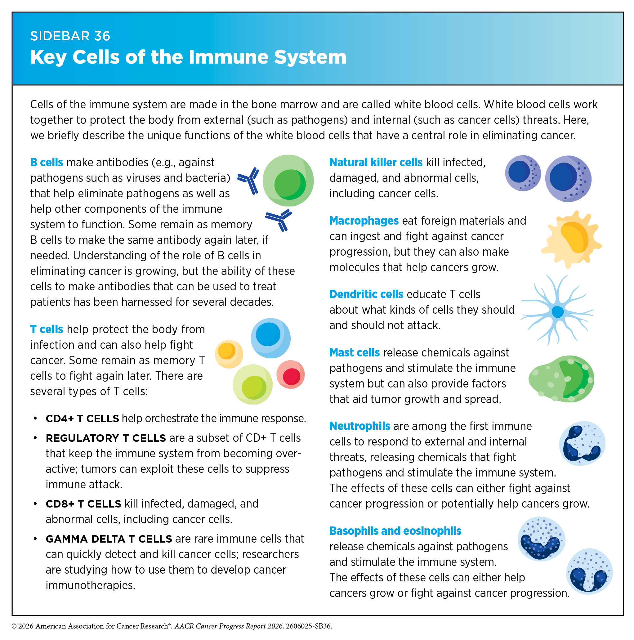
The immune system has two main components. The innate immune system includes physical barriers, such as the skin and mucous membranes, as well as certain immune cells and molecules that respond to and kill a broad range of pathogens. In contrast, the adaptive immune system provides a specific response against pathogens and remembers them for a faster response in case of a future encounter. Some key immune cells include dendritic cells that detect threats and alert the immune system to respond, T cells that can kill infected or abnormal cells and help coordinate the immune response, and B cells that produce antibodies to neutralize pathogens (see Sidebar 36).
Although many abnormal cells are eliminated by the immune system, as cancer progresses, some cancer cells acquire properties that allow them to evade immune detection and destruction. Researchers have identified several mechanisms by which these cells evade the immune system, including activating immune checkpoints that suppress T-cell responses, recruiting immunosuppressive cells, and releasing proteins and signaling molecules that impair immune cell function (158)Tufail M, et al. (2025) Signal Transduct Target Ther, 10: 227..
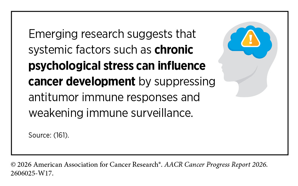
In other cases, cancer cells can alter signaling pathways and metabolic interactions within the TME to suppress immune responses. A recent study found that tumor-associated macrophages, a type of immune cell, can provide metabolic support that helps glioblastoma cells suppress immune responses (159)Li D, et al. (2026) Nat Cell Biol, 28: 349.. In another study, researchers showed that cancer cells can hijack mitochondria—the energy-producing structures inside cells—from immune cells, impairing immune response and helping tumors spread to nearby lymph nodes (160)Terasaki A, et al. (2026) Cell Metab, 38: 388..
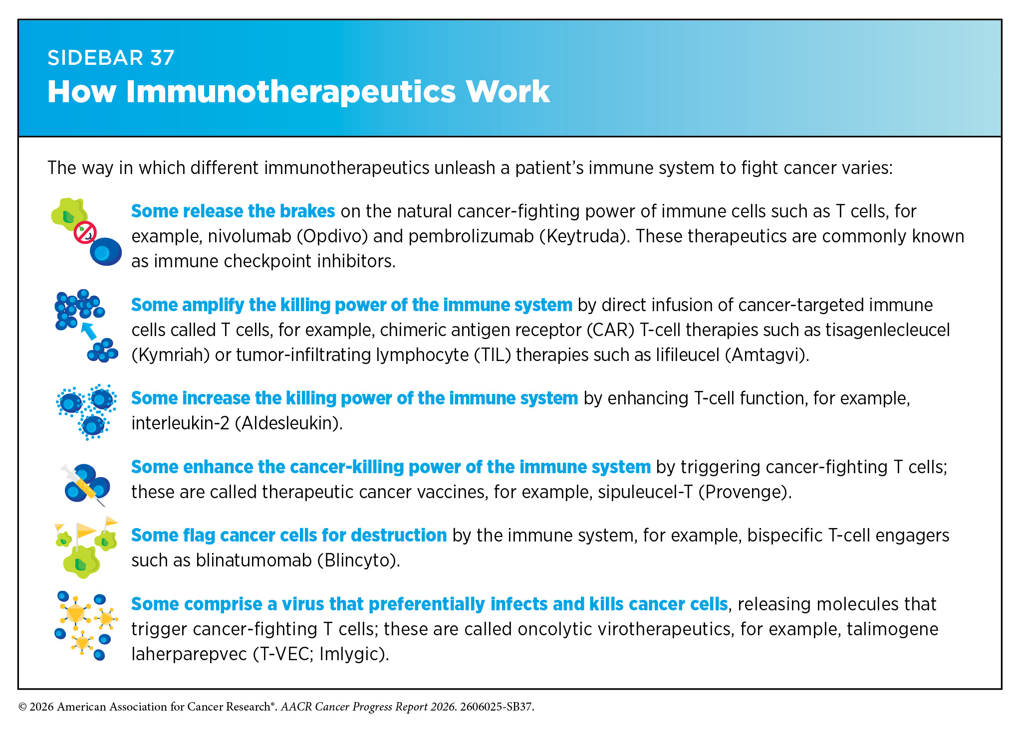
Researchers continue to explore therapeutics that target immune escape mechanisms. ICIs, for example, block proteins that tumors use to suppress T-cell activity, unleashing the immune response (see Sidebar 37). In a recent study, researchers identified a previously unrecognized immune checkpoint that limits T-cell activity within tumors. They found that selective serotonin reuptake inhibitors (SSRIs), a class of antidepressant drugs, enhanced T-cell antitumor activity and improved responses to ICI therapy in preclinical models by blocking this pathway, highlighting the potential to repurpose existing drugs to improve cancer immunotherapy (162)Li B, et al. (2025) Cell, 188: 3823.. As our understanding of the complex interactions between cancer and the immune system continues to grow, so does the potential to develop more effective and lasting therapies.
Nervous System
The nervous system—composed of the CNS, which includes the brain and spinal cord, and the peripheral nervous system (PNS), a network of nerves that connects the CNS to the rest of the body—uses electrical signals and chemicals to regulate and coordinate essential functions. Recently, the nervous system has become more recognized as a key regulator of cancer development not only in brain tumors but also across cancer types, including pancreatic, breast, stomach, colorectal, lung, and prostate cancers (163)Monje M, et al. (2025) Nat Cancer, 6: 1928..
Research has uncovered dynamic interactions between the nervous system, the immune system, and cancer as drivers of tumor growth, metastasis, immune evasion, and resistance to therapy, giving rise to the emerging interdisciplinary field of cancer neuroscience. These interactions take place both within the local TME and systemically throughout the body.
Nerve cells, or neurons, interact with cancer and non-cancer cells by releasing signaling molecules and transmitting electrical signals through direct contact within the TME. Recent studies have shown that tumors can actively remodel nearby nerves and promote cancer development. For example, using single-cell and transcriptomic technologies (see Sidebar 8), researchers mapped the cellular interactions surrounding nerves in pancreatic tumors and found that cancer cells reprogram nearby nerve-associated cells and reshape local stromal and immune environments, creating a local environment that supports tumor progression (164)Chen MM, et al. (2025) Cancer Cell, 43: 1656..
Research has found that lung cancer cells communicate with the nervous system in multiple ways. Two recent studies showed that small cell lung cancer cells can interact with nearby peripheral nerves through neurotransmitter signaling, including by forming functional neuron–tumor connections that promote tumor growth (165)Savchuk S, et al. (2025) Nature, 646: 1232.(166)Sakthivelu V, et al. (2025) Nature, 646: 1243.. In another study, researchers showed that lung tumors can also communicate with the brain through sensory nerves, triggering signals from the brain back to the tumor that suppress immune responses and promote tumor growth (167)Wei HK, et al. (2026) Nature, 650: 1007.. In all three studies, disrupting these neural interactions reduced tumor growth in preclinical models, highlighting cancer–nerve communication as a potential therapeutic target.
Researchers have also uncovered important roles for the nervous system in regulating antitumor immunity. In stomach cancer, researchers found that neural signaling reprogrammed tumor metabolism, altered communication with immune cells, and promoted an immunosuppressive environment (168)Liao F, et al. (2026) Cancer Res, 86: 1968.. In another study, researchers showed that cancer-induced nerve damage can trigger an inflammatory repair response that was associated with resistance to immunotherapy across multiple cancer types (169)Baruch EN, et al. (2025) Nature, 646: 462..
Scientists are discovering new ways in which the nervous system influences cancer progression. Factors such as nerve activity, brain chemical signaling, and disruption to the body’s internal clock are being explored for their roles in promoting cancer cell proliferation, survival, and immune evasion.
Microbiome
The microbiome is made up of all the microbes—bacteria, fungi, viruses, and other microorganisms—that live on and in the skin, mouth, gut, and other sites in the body. Studies have shown that microbiomes play essential roles in digestion, metabolism, immune system regulation, and protection against pathogens. A growing body of research suggests that these microbial communities also influence cancer development and treatment response by shaping immune activity, inflammation, and the TME (91)Hanahan D (2026) Cell, 189: 2254.. While many early studies focused on the gut microbiome, scientists are now recognizing the importance of microbiomes in other tissues and organs, each with its own distinct microbial communities (170)Yao Y, et al. (2026) Signal Transduct Target Ther, 11: 39..
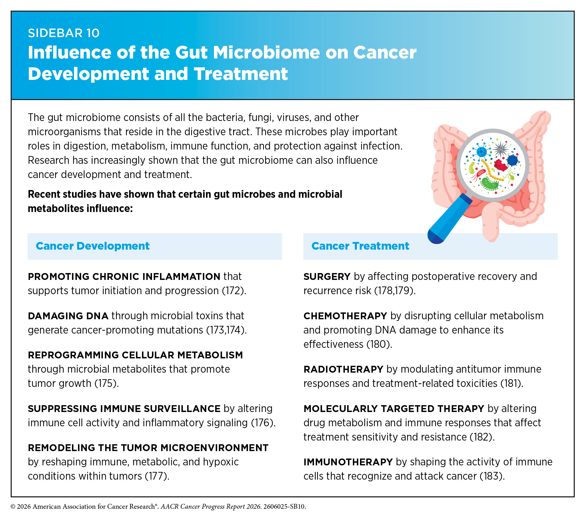
Research has shown that changes in the composition of the gut microbiome can contribute to the initiation and progression of colorectal, stomach, liver, pancreatic, breast, and prostate cancers (171)Nobels A, et al. (2025) Nat Metab, 7: 895.. Beyond the gut microbiome’s role in cancer development, certain gut microbes and microbial metabolites can also affect response to cancer treatment (see Sidebar 10).
Recent studies have shown that modulating the gut microbiome may improve the effectiveness of cancer immunotherapies (see Releasing the Brakes on the Immune System). One approach is fecal microbiota transplantation (FMT), a procedure in which stool from a donor is transferred to a patient to introduce beneficial microbes to the gut. Three landmark clinical trials recently demonstrated the promise of this approach. In patients with melanoma, kidney, and lung cancers, FMT was combined with immune checkpoint inhibitor therapy and showed encouraging signs of improving responses to treatment (184)Porcari S, et al. (2026) Nat Med, 32: 1316.(185)Fernandes R, et al. (2026) Nat Med, 32: 1325.(186)Duttagupta S, et al. (2026) Nat Med, 32: 1337.. Other research suggests that microbiome modulation may also improve chimeric antigen receptor (CAR) T-cell therapies. For example, a recent study found that supplementing with specific bacterial species enhanced the effectiveness of CAR T-cell therapy in preclinical models (187)Marcos-Kovandzic L, et al. (2025) Cancer Discov, 15: 1905..
Beyond the gut, researchers have found that the oral microbiome may contribute to cancer development in distant organs. Certain oral bacteria have been identified within breast tumors that promote cancer tumor growth and metastasis by inducing inflammation and DNA damage (188)Parida S, et al. (2026) Cell Commun Signal, 24.. However, more research is needed to further characterize the microbiome inside tumors and determine their influence on cancer development.
It is becoming increasingly clear that targeting the microbiome may help improve health outcomes for patients with cancer (189)Hajjar R, et al. (2026) Nat Rev Microbiol, 24: 392.. Efforts are now underway to manipulate the microbiome through strategies such as specifically designed mixtures of beneficial bacteria (probiotics), dietary supplements that nourish helpful microbes (precision prebiotics), and engineered bacterial species that deliver therapies or modulate immune signaling.
Tumor Microenvironment
The TME—composed of cancer and supportive, non-cancer cells, blood vessels, signaling molecules, and structural components—is a dynamic biological system that influences tumor growth, metastasis, and response to treatment. Cancer cells shape this environment by releasing molecules that secure nutrients, oxygen, and structural support essential for their growth (see Sidebar 9).
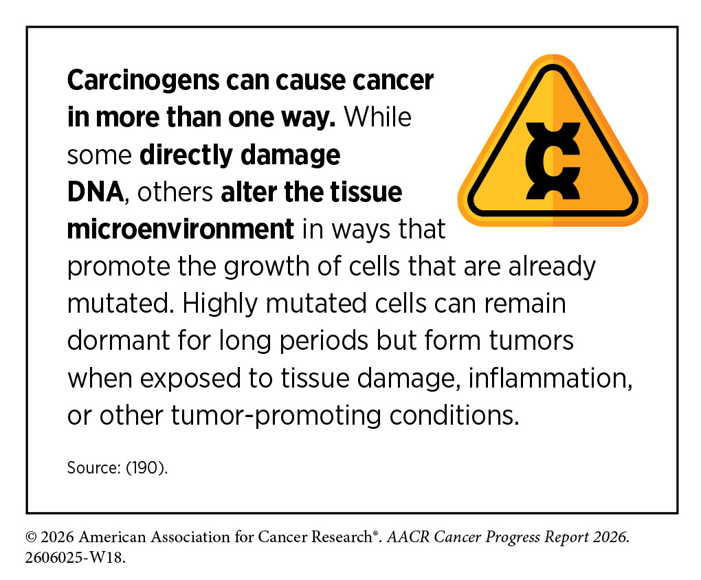
Understanding the TME is essential for gaining insights into cancer development and advancing cancer therapies, as it reveals how the surrounding cellular and structural environment supports tumor growth and may be targeted to improve therapeutic outcomes.
Recent advances in single-cell and spatial transcriptomic technologies (see Sidebar 8) are transforming our understanding of the TME by helping map the cellular and molecular interactions that occur within and around tumors. These technologies can determine how cells organize and communicate across three dimensions within the microenvironment, uncovering evolving processes that promote or suppress tumor growth and metastasis, as well as influence responses to therapy. In a recent study, researchers used multi-omic, single-cell, and spatial profiling to reveal how mutations in the TP53 gene remodel the TME in lung cancer. They found that these mutations remodel the TME by promoting interactions among cancer cells, immune cells, and stromal cells that support tumor progression and metastasis, while also increasing immune checkpoint signaling that may make tumors more responsive to immunotherapy (191)Zhao W, et al. (2025) Nat Cancer, 6: 1857..
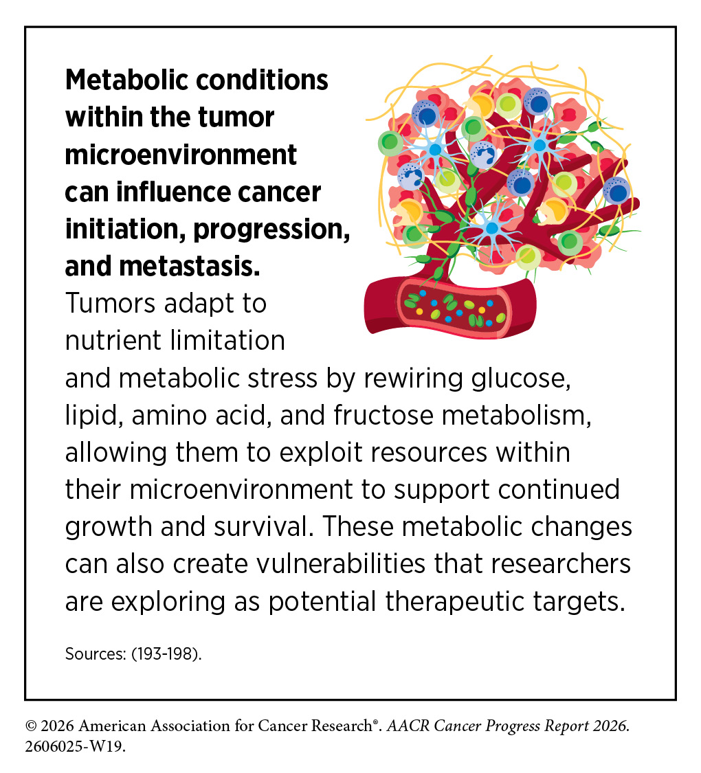
Studying the TME in cancers that metastasize to the brain may offer valuable insights into why these tumors are often difficult to treat. Beyond the challenge of delivering drugs across the blood–brain barrier, the brain microenvironment itself can suppress immune responses and provide signals that help metastatic cancer cells survive and resist therapy. To better understand this environment, researchers are using advanced technologies that analyze individual cells and their activity within the tumor’s structure. In a recent study, researchers built a comprehensive single-cell atlas of brain metastases across several cancer types, revealing features that help tumors adapt and survive in the brain microenvironment (192)Xing X, et al. (2025) Cancer Cell, 43: 1242.. These findings highlight how studying tumor cells and their surroundings can reveal molecular underpinnings of tumor growth and resistance to treatment and may point to new ways to improve therapy.
Normal aging is the greatest risk factor for cancer. As tissues age, changes in the TME—including declining immune surveillance, chronic inflammation, and cellular senescence—create tumor-promoting conditions, although some age-related changes have context-dependent effects on cancer initiation and progression. For example, one recent study found that aging can promote metastatic spread by activating stress response pathways that increased lung tumor plasticity and adaptability (199)Patel AAH, et al. (2026) Nature, 652: 1339.. In contrast, additional studies have shown that aging altered the lung microenvironment in ways that reduced the initiation of KRAS-driven lung tumors and also slowed the growth of tumors that did develop by reshaping both cancer cells and their surrounding microenvironment (200)Shuldiner EG, et al. (2025) Nat Aging, 5: 2263.(201)Zhuang X, et al. (2025) Nature, 637: 184.. Together, these findings highlight how age-related changes in the TME can have both tumor-promoting and tumor-suppressive effects that influence cancer development.
Within the TME, the tumor immune microenvironment (TIME) refers to the network of immune cells and immune-modulating factors that interact with the tumor. The TIME plays an important role in cancer development and response to treatment, and a deeper understanding of these processes is essential for advancing effective immunotherapies.
Emerging research suggests that age-related weakening of the immune system, known as immune aging, makes it harder for the body to detect and eliminate cancer cells and may alter the TIME in ways that support tumor growth and treatment resistance. For example, research has found that age-associated clonal hematopoiesis may contribute to these changes by altering the composition and behavior of tumor-infiltrating immune cells, remodeling the TIME in ways that promote tumor progression and are associated with poorer clinical outcomes (202)Pich O, et al. (2025) N Engl J Med, 392: 1594..
Integrating Knowledge to Advance Cancer Research and Precision Medicine
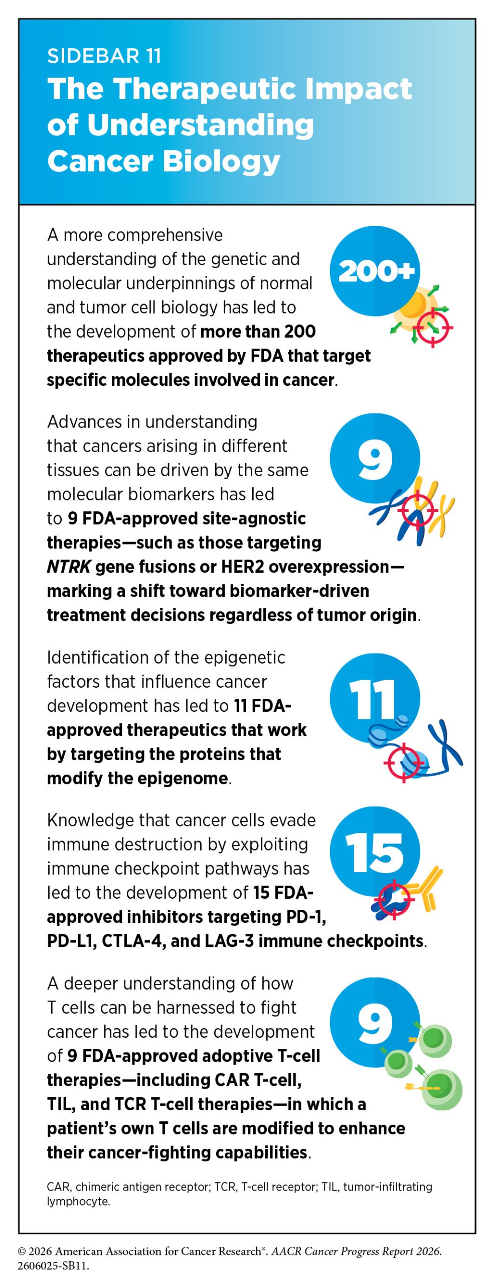
Breakthrough discoveries and technological innovations have significantly advanced our understanding of cancer initiation and progression, enabling the development of a myriad of effective anticancer therapies in recent years (see Sidebar 11). A crucial insight stemming from this knowledge is the understanding that each patient’s cancer is unique.
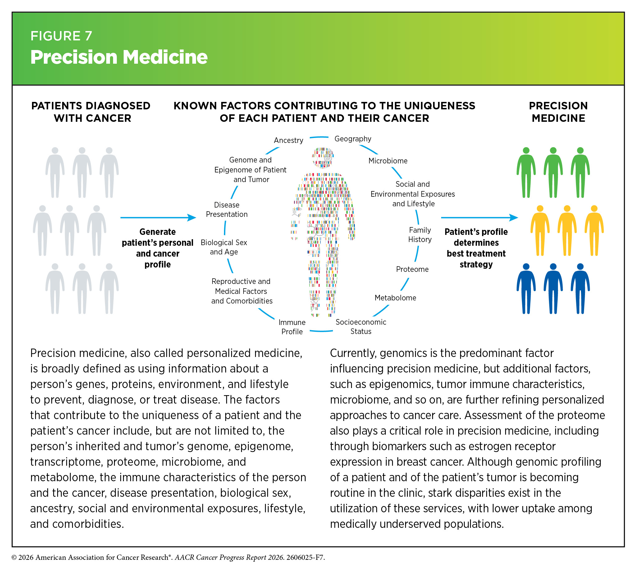
This understanding has paved the way for precision medicine, also called personalized medicine, which is broadly defined as using information about a person’s genes, proteins, environment, and lifestyle to prevent, diagnose, or treat disease (see Figure 7). For patients with cancer, precision medicine means using molecular characteristics of the tumor, such as the genome sequence of cancer cells or features of the tumor-associated immune cells, to make a diagnosis, plan treatment, and predict and monitor treatment response.
Technological Innovations and Collaborative Science
By leveraging emerging technologies and multi-omic data (see Sidebar 8), researchers are generating a complete picture of the cellular and molecular characteristics that drive cancer. This comprehensive approach, coupled with powerful computational tools and shared research platforms, is enabling deeper integration of cancer biology insights and driving the development of more personalized therapies, refining tumor classification, revealing novel therapeutic targets, and addressing key challenges such as treatment resistance. For example, by integrating whole-genome sequencing with complementary molecular analyses and clinical data, researchers can identify clinically actionable biomarkers that may be missed by more limited testing approaches and can inform diagnosis, hereditary cancer risk assessment, and treatment selection (204)van Putten J, et al. (2026) Nat Med, 32: 1286.(205)Kim R, et al. (2026) Nature, 649: 1282..
These advances are increasingly being implemented through large-scale precision medicine initiatives that integrate genomic and clinical data across populations. National programs such as Germany’s genomDE strategy, Finland’s FinnGen project, and the Taiwan Precision Medicine Initiative are creating extensive genomic and health data resources that support biomarker identification, risk prediction, and the development of more personalized approaches to prevention, diagnosis, and treatment (206)Till A, et al. (2025) Nat Med, 31: 3981.(207)Aavikko M, et al. (2026) Nat Rev Cancer, 26: 159.(208)Yang HC, et al. (2025) Nature, 648: 117..
AI and advanced computational approaches are becoming essential tools for translating the growing volume of biological and clinical data into actionable insights that can guide precision oncology. Recent advances have highlighted the ability of AI-driven approaches to integrate diverse datasets, improve patient stratification, predict treatment response, and accelerate the development of new therapies (209)Liu F, et al. (2026) Nat Rev Cancer, 26: 497.(210)Goodwin RJA, et al. (2026) Cancer Discov, 16: 847.. Reflecting the increasing importance of AI-driven approaches in cancer research, an executive order issued in September 2025 directed federal agencies to expand the use of AI in pediatric cancer research through the Childhood Cancer Data Initiative. This order emphasized leveraging large-scale health and research data resources to improve cancer diagnosis, biomarker discovery, clinical trial design, treatment development, and prevention strategies (see Improving the Use of Digital Information in Cancer Treatment and Management) (211)The White House. Unlocking Cures for Pediatric Cancer with Artificial Intelligence. Accessed: June 30, 2026..
Large-scale data-sharing initiatives, such as The Cancer Genome Atlas (TCGA) and the Cancer Dependency Map (DepMap), have laid the groundwork for a more comprehensive understanding of cancer biology. TCGA has enabled an extensive catalog of genetic, epigenetic, and molecular changes, or “maps,” across patient tumors, thus providing a reference point for distinguishing tumor subtypes and identifying common oncogenic pathways. DepMap has complemented this effort by providing a large-scale data repository for genetic and molecular vulnerabilities, or weak points that cancer cells rely on for survival, across a wide array of cancer models routinely used by researchers (see Sidebar 4). In April 2026, DepMap announced the launch of its next phase, which aims to accelerate the discovery of emerging therapeutic modalities while advancing efforts to overcome therapeutic resistance (213)Broad Institute. Cancer Dependency Map Consortium launches Phase 3 to accelerate next-generation therapeutics. Accessed: June 30, 2026..
Building on these foundational resources, the Human Tumor Atlas Network (HTAN), a collaborative, data-intensive cancer research initiative launched as part of the Cancer Moonshot, is applying advanced technologies to study individual cells and their molecular features within the structure of tumors. HTAN studies use single-cell, spatial, and multi-omic technologies (see Sidebar 8, p.41)—including genomic, transcriptomic, proteomic, and advanced imaging approaches—to map the cellular and molecular architecture of tumors and premalignant tissues across disease initiation, progression, and metastasis (214)Enninful A, et al. (2026) Nat Methods.(215)Jiménez-Sánchez A, et al. (2026) Cancer Res, 86: 1769.(216)Tandukar B, et al. (2026) Nat Cell Biol, 28: 1300.(217)Tandukar B, et al. (2025) Nat Commun, 16: 10663.. As of June 2026, HTAN investigators have generated 14 tumor atlases spanning 20 organ sites, providing a resource empowering researchers across the globe to decode the cellular and molecular complexity of cancer at unprecedented resolution (218)National Cancer Institute. NCI Human Tumor Atlas Network. Accessed: June 30, 2026..
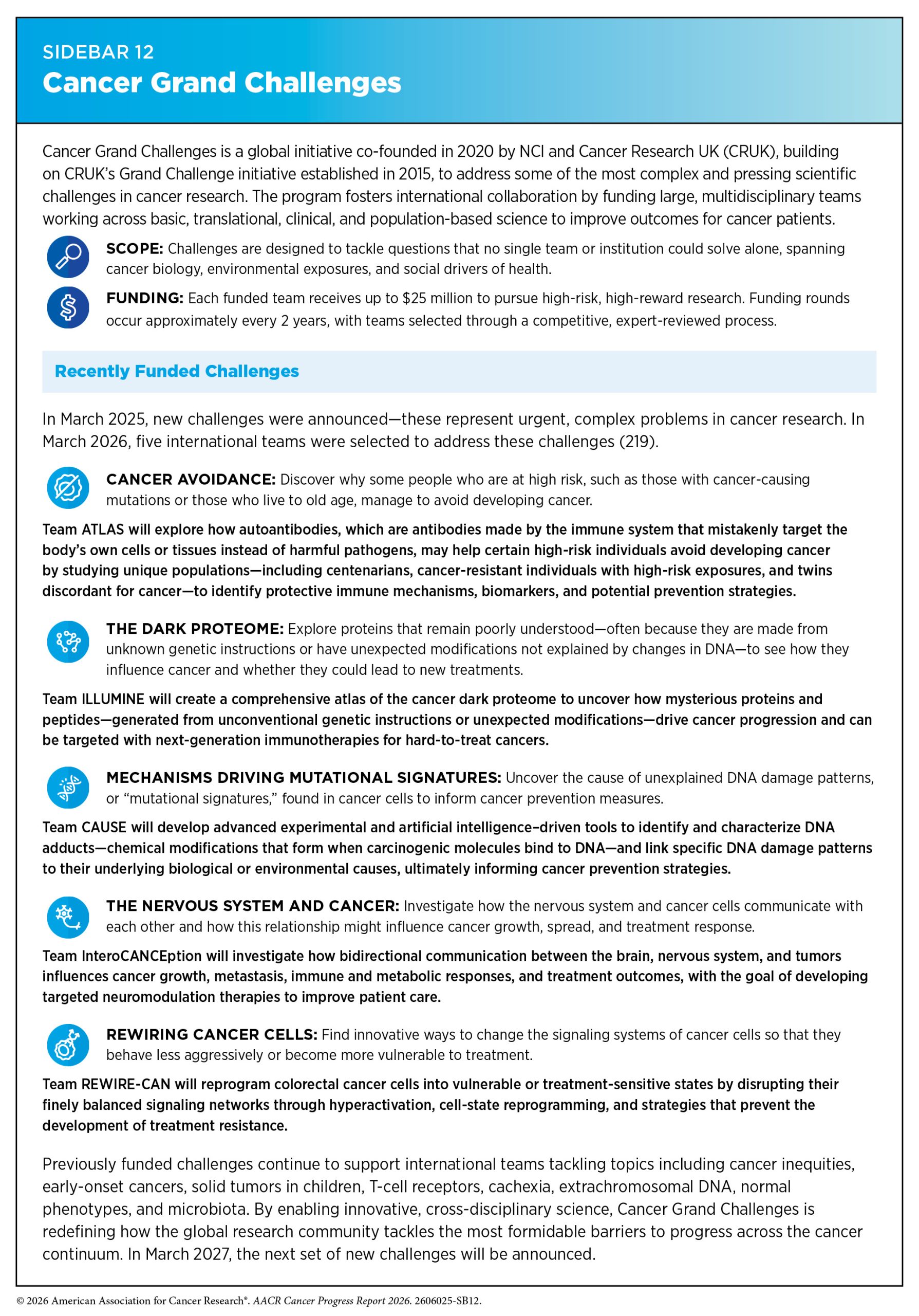
Beyond generating data resources, international collaborative research initiatives, such as the Cancer Grand Challenges, are applying integrative approaches to tackle some of cancer’s most complex and unresolved questions. Launched as a global partnership between the National Cancer Institute and Cancer Research UK, Cancer Grand Challenges brings together multidisciplinary teams from around the world to tackle scientific challenges that are too complex for any single laboratory or institution to address (see Sidebar 12).
These challenges focus on critical unmet needs that demand integrated research spanning cancer biology, environmental exposures, and social drivers of health, such as determining the cause of early-onset cancers; understanding the biology of ecDNA and the dark proteome; addressing cancer cachexia (see Challenges Faced by Survivors); developing novel therapies for pediatric cancers; addressing cancer inequities; and uncovering the dynamic interactions between the nervous system and cancer. By supporting bold, collaborative science that spans basic, translational, clinical, and population-based research, this initiative highlights the importance of integrating knowledge across domains to advance cancer prevention, detection, and treatment for all patients.
As precision oncology advances, it is essential to ensure that its benefits are accessible to all patients. Researchers have recently emphasized the need to diversify genomic datasets and include ancestral and environmental variables in both research and clinical implementation (220)Liu Y, et al. (2026) Cancer Discov, 16: 660.. Moving forward, broadening representation in studies and making molecular testing and new treatments more accessible across patient populations will be essential to realizing the full potential of precision medicine for all patients (see Progress Across the Clinical Cancer Care Continuum).
Next Section: Reducing the Risk of Cancer Development Previous Section: Cancer in 2026